What are the different types of MRI scan we used?
Localizer scan - this very short scan (12 seconds) allows the scanner to work out exactly where your brain is inside the magnet.
T1-weighted scan - the first long scan and the most important structural scan for our research. It gives a clear picture of what the brain looks like and so is used to measure the thickness, surface area, and volume of cortex.(For more information on brain structure see question 9.) This scan uses the differences in the amount of water and fat that different brain tissues contain (e.g., grey- vs white-matter) to produce differences in the brightness of the image. “T1-weighted” refers to the way in which the measurement of the brain’s magnetic field is done to favour tissues containing less water and more fat such as white-matter, so that they appear brighter in the final picture than grey-matter or the fluid that the brain floats around in. Click here for more information on how MRI works.
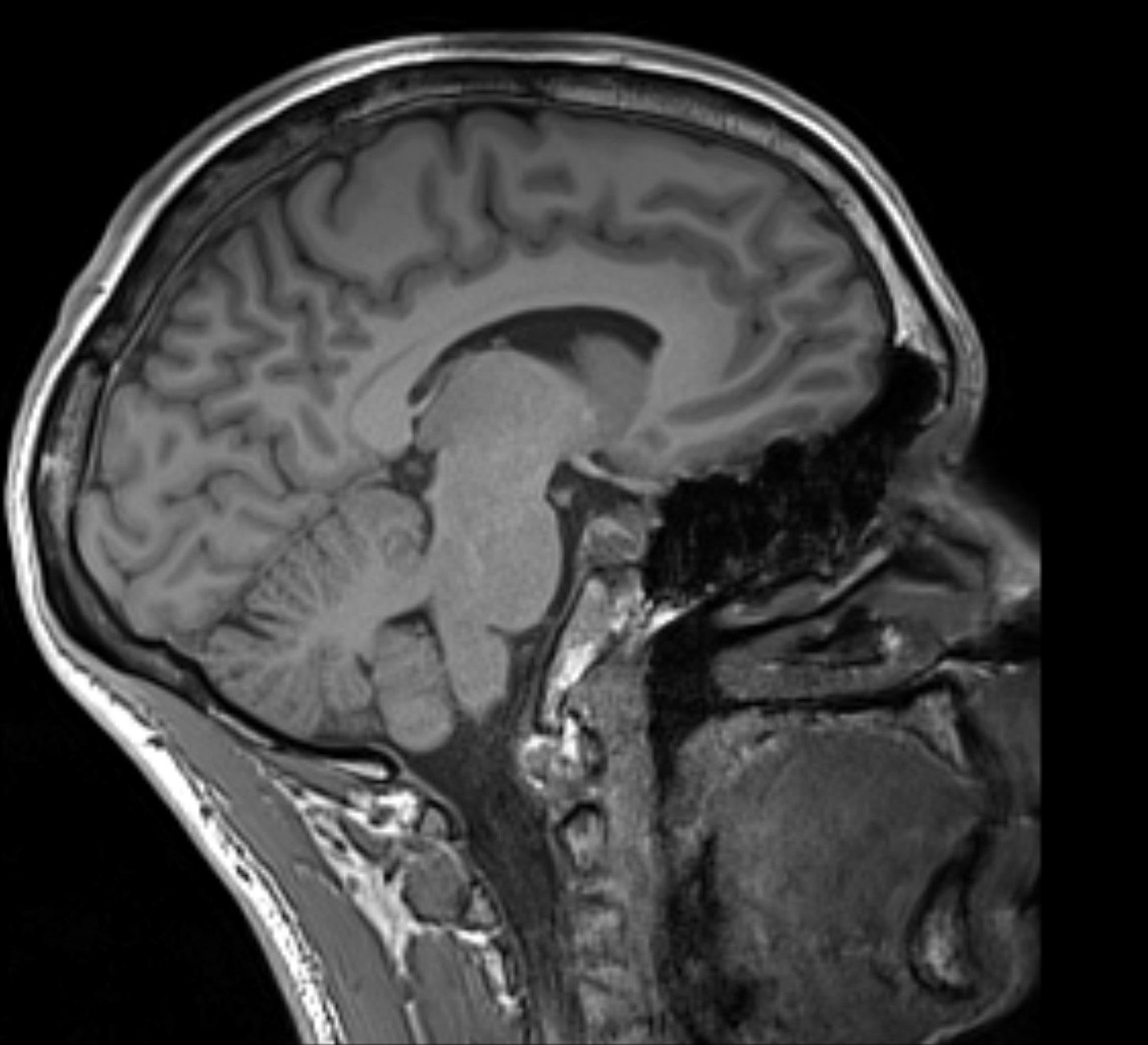
Image from a T1-weighted scan showing grey-matter darker than white-matter.
Field map - used to measure the strength of the main magnetic field in the scanner tube at the point where the head was. This main magnetic field needs to be exactly the same strength at all points in this area but will not be when a person is inside the scanner. Once this scan has mapped out the magnetic field strength, this information can be used later to correct for any distortion in the data.
Resting-state scan - the long one without music where we asked Study members to keep their eyes open – this was used to get an idea of what the brain was doing when it was not actively engaged in one of our tasks. Even though we normally tend to think of our brains being more active when we are concentrating on something, there are some networks (parts of the brain that are connected and become active at the same time) in the brain that actually chatter to each-other more when you are not focussing on a task. That’s why we asked Study members to not think about anything in particular and to stare at a boring grey screen. Although Study members would not have realised this at the time, it is actually the same type of scan as used during the fMRI (functional MRI) games, just not one where they were asked to do a task.
fMRI Games - during each of these scans, images and words were shown to Study members on a computer screen, and they were asked to respond in various ways using buttons that they could press with one hand. As they concentrated on the tasks, the scanner measured how active different parts of their brain were (Click here for more information on how this works):
- Matching Game - Study members were asked to match either shapes, or faces with different emotional expressions, which can help us better understand how we may react to stress or danger.
- Colours Game - this game is better known in psychology as a Stroop Test. Study members were shown the names of colours and asked to identify the colour in which the words were written. The challenge was that the names of the colours and the colour in which they were written were not always the same (e.g.,"YELLOW"). This game challenges our ability to ignore one type of information in favour of another, which is helpful in making decisions.
- Quickstrike - in this game, Study members were asked to press a button as quickly as they could when they saw a particular symbol. If they pressed it fast enough they won an amount of money which was paid to them after the scans. This game helps us understand how sensitive we may be to rewards and how motivated we may be to chase after our goals.
- Faces Game - this involved trying to memorize a series of faces and names and then recalling them later in the game. This was a very difficult task but it helps us better understand how well we can remember new information.
DWI (Diffusion-weighted imaging) - Study members will probably remember this one because the scanner bed vibrated (see question 5). It measures how easily water molecules can randomly diffuse (move) in the brain’s white-matter (see question 9). Water molecules can move more easily in the same direction that nerve fibres run in than across them (imagine dragging a fork in different directions through cooked spaghetti). By comparing the movement of water in different directions we can get information about how well different parts of the brain are connected. These scans produce the lovely coloured picture that we sent to Study members later, in which the bundles of nerve fibres running in different directions are colour-coded during the analysis process.
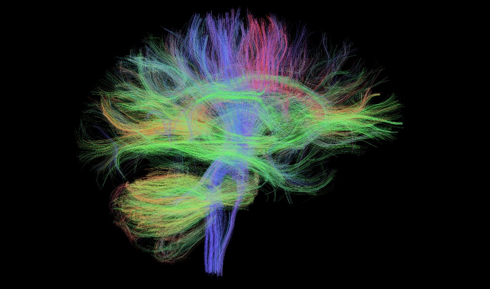
Image from a DWI scan showing white-matter nerve fibre pathways. They have been colour-coded during analysis to show their directions of travel.
SWI (Susceptibility-Weighted Imaging) - in this scan Study members were just listening to the music and staying still. We did this scan to check for any “dark spots” - areas of blood break-down products in the brain that could have come from damaged small blood vessels. The basic way in which it does this is similar to fMRI i.e., blood changes the strength of the magnetic field due to the iron in red blood cells (for more information click here ). “Susceptibility” just refers to how much something becomes magnetized when it is put into a magnetic field.
T2-weighted scan - the very last scan of the session by which time all our Study members could think about was getting out of the tube! As with the T1-weighted scan at the beginning, this scan used the differences in the amount of water and fat that different brain tissues contain (for more information click here). We used this scan to produce particularly good contrast between the white-matter which shows up as dark (the opposite of the T1 scan) and “bright spots” called White-Matter Hyperintensities (WMHs) which contain more water than the surrounding white-matter (for more information see question 12).
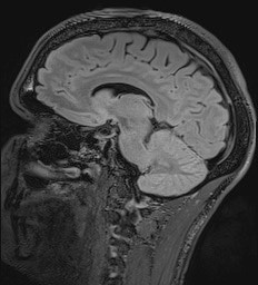
Image from a T2-weighted scan with grey-matter lighter than white-matter.
Click here to return to the MRI Q&A page