Retinal Imaging shows microvascular abnormality in schizophrenia
Sunday 1st December 2013
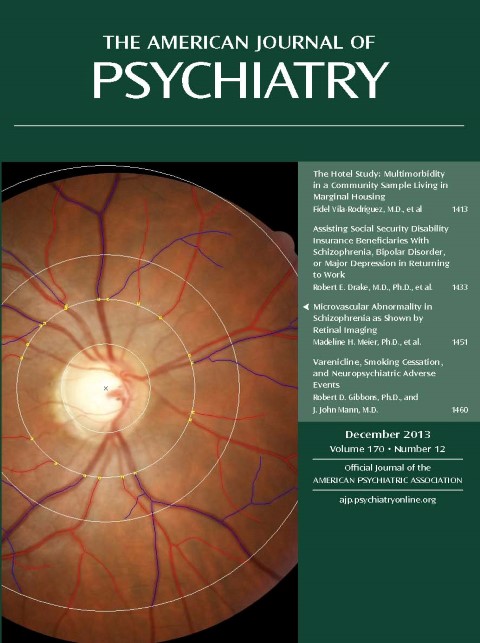
At the age 38 assessment, the Dunedin Study introduced retinal imaging as part of the assessment. Using these data, a recent study showed that those who developed schizophrenia were distinguished by wider retinal venules, suggesting microvascular abnormality reflective of insufficient brain oxygen supply.
The wider retinal venules were not simply an artifact of co-occurring health problems in schizophrenia patients. Wider venules were also associated with a dimensional measure of adult psychosis symptoms and with psychosis symptoms reported in childhood. The findings provide initial support for the hypothesis that individuals with schizophrenia show microvascular abnormality. Moreover, the results suggest that the same vascular mechanisms underlie subthreshold symptoms and clinical disorder and that these associations may begin early in life.
Meier, M.H., Shalev, I., Moffitt, T.E., Kapur, S., Keefe, R., Wong, T.Y., Belsky, D.W., Harrington, H. L., Hogan, S., Houts, R., Caspi, A. & Poulton, R. | 2013
Microvascular Abnormality in Schizophrenia as Shown by Retinal Imaging
American Journal of Psychiatry, 2013, 170(170), 1451-1459.
http://ajp.psychiatryonline.org/article.aspx?articleid=1738034