How does MRI work?
Most of the atoms in your body are hydrogen atoms - in particular water and fat contain large numbers of them. Every atom has a bit in the middle called a nucleus and in the case of hydrogen the nucleus is just a single positively-charged particle called a proton. Here’s the really important thing – a tiny particle such as a proton behaves in some ways a bit like a little bar magnet with two poles that has a fundamental property called “spin” (although confusingly “spin” is a mathematical construct – the proton doesn’t actually spin around or at least not in the way that we would normally think about). Despite this, the property of spin means that when they are put into the scanner’s main magnet, the hydrogen protons in the body part being scanned (in your case your brain) behave like wobbling children’s tops, rotating around at an angle at millions of times per second. During a scan some of the other magnets in the scanner walls rapidly oscillate (increase and decrease) the strength of their magnetic field at a very specific frequency (in fact, it has to be the same frequency as the protons are rotating). The hydrogen protons absorb energy from this oscillating magnetic field which causes them to develop a slight overall synchronization with each other. This is what resonance is - the same basic idea as pushing a child on a swing – if you pass energy to the swing (i.e., push it) with the right timing (i.e., the same frequency that the swing is already going at) it gets higher and higher. The nett effect of these mildly synchronized hydrogen protons is a rotating magnetic field which can be measured by antennas in the scanner or in the special helmet that Study members wore. When the scanner’s oscillating magnetic field then switches off, the hydrogen protons return to their previous unsynchronized state, in the process releasing energy, and the brain’s rotating magnetic field declines back to zero (like stopping pushing a swing – the swing loses energy to air friction, friction of the rope on the branch, etc. and comes to a stop).
Very importantly, different types of brain tissue have different amounts of water and fat in them which means that the energy is released and the brain’s magnetic field declines at different rates depending on the tissue. MRI takes advantage of this to produce pictures with contrasting lightness and darkness for different tissues. So for example, in the main structural scan that we used (T1-weighted scan), the fluid that your brain floats around in which is 99% water, would show up as darker than the white matter which contains nerve fibres wrapped in fat. In a different scan type like the T2-weighted scan this would be reversed and the white matter would be darker than the fluid. Click here for more details on the different types of scan.
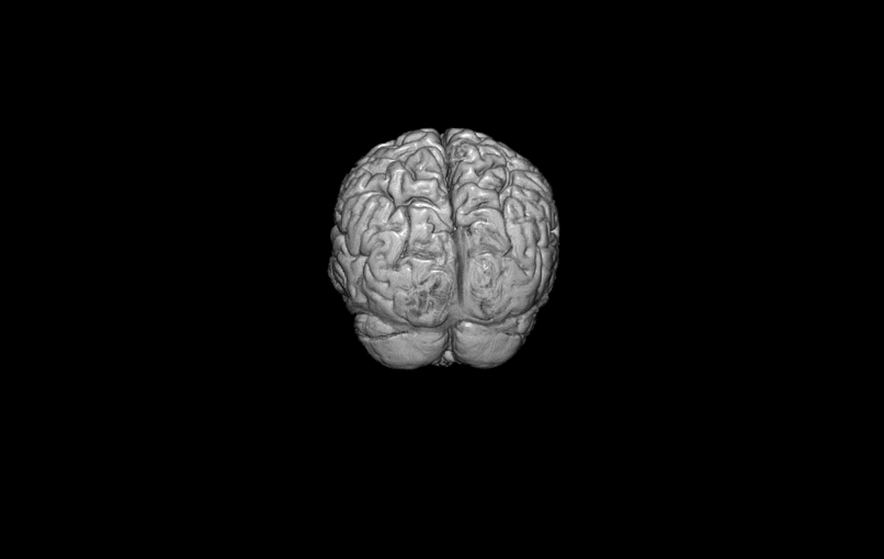
In general MRI scans take virtual “slices” of the brain, usually millimetres thick, and each slice is divided up into little areas only millimetres across. The end result is a division of the brain up into tiny 3D cubes a bit like the way that a computer screen is divided into pixels. But in this case they are 3-dimensional and so are called voxels (“volume pixels”). The pictures of the brain that we see are formed by adding together all these voxels. Because they are 3-dimensional the voxels can also be put together to make 3D images of the brain like the ones that we sent to Study members after their scans. This is also how we measure the volume of different parts of the brain. The dividing up into slices and bits of slices is done using a different set of scanner magnets that switch on and off as needed and cause the overall magnetic field in the scanner to vary. This is the cause of the loud noises that the scanner makes.
(1) “An animation showing the thin cross-sections (“slices”) of the brain that the MRI scanner makes.”
(2) “The virtual slices can be used to create a 3D image of the brain”